GUT MICROBIOTA AND IT’S ROLE IN OBESITY

INTRODUCTION

Obesity is becoming the most dangerous lifestyle disease of our time, and its effects are already being observed in both developed and developing countries. Obesity can be described as an increase in the adipose tissue that releases a wide variety of proinflammatory cytokines and chemokines, called adipokines, promoting inflammation, recruitment of macrophages and insulin resistance. Multiple etiological factors have been attributed to the genesis of obesity, of which hereditary predisposition, wrong dietary habits and life-style (lack of exercise) , besides change in the gut microbiome composition also play another vital role in genesis of obesity.

Human gut is the habitat of diverse and dynamic microbial ecosystems. The collection of bacteria, archaea and fungi inhabiting in the human gastrointestinal tract is termed as ‘Gut microbiota’. A biological mutualism relationship exist between gut microbiota and host. The number of microorganisms inhabiting the GI tract has been estimated to exceed 1014, which encompasses ∼10 times more bacterial cells than the number of human cells and over 100 times the amount of genomic content (microbiome) as the human genome.

The microbiota offers many benefits to the host, through a range of physiological functions such as

  • Strengthening gut integrity or shaping the intestinal epithelium
  • Regulating host immunity
  • Produce substances that are antifungal, antiviral and antibacterial & reduce the risk of infections.
  • Produce substances such as antidiabetic, anticancer, antiobesity, stress(mental and physical), anti asthma, anti chronic pulmonary
  • Strengthen intestinal barrier system & prevent allergy and hypersensitivity reactions.

However, there is potential for these mechanisms to be disrupted as a result of an altered microbial composition, known as dysbiosis(or an imbalance between commensal and pathogenic organisms). Healthy microbiome, can vary distinctly between gender, across different ages, geographical regions and medical contexts.

Recently, it has been observed that the composition of gut microbiota of healthy persons is different from that of obese ones. Such observations suggested a possible relationship between the compositional pattern of gut microbiota & pathology of metabolic disorders. Currently, advanced technique for bacterial DNA sequencing & bioinformatics tools have contributing to identify bacterial communities in various orders. Phyla constitute the highest level of classification, which is sequentially subdivided into class, order, family, genus and species. The most predominant phyla (90%) in human gut microbiota are phyla, Bacteroidetes (Gram negative), Firmicutes (Gram positive) and Actinobacteria (Gram positive), are most abundant and have been found to play a dominant role in the pathophysiology of metabolic disorders specifically, obesity. Other phyla also contribute, but to a lesser degree.

DEVELOPMENT OF HUMAN GI MICROBIOTA

The composition of gut microbiota is unique to each individual, just like our fingerprints. Composition of gut microbiota starts to define each individual from beginning  of the birth, through several factors such as mode of delivery (caesarean delivery vs. vaginal delivery), breast milk vs. formula feeding, antibiotic usage, and timing of the introduction of solid foods and diet.

MODE OF DELIVERY

For vaginally born infants, Lactobacillus (phyla Firmicutes) dominates the gut, but within months there is a greater distribution of Bifidobacterium (phyla Actinobacteria) and Bacteroides (phyla Bacteroidetes) microbiota composition. High abundance of lactobacilli during the first few days, is due to a reflection of the high load of lactobacilli in the vaginal flora. Within the Lactobacillus genus, different species are associated with an obese profile (Lactobacillus reuteri) or a lean profile (Lactobacillus gasseri and Lactobacillus plantarum), such that the microbiota composition is related to body weight and obesity at the species level.

In contrast, the gut microbiome of an infant born by caesarean section are comprised of a bacteria that are transferred horizontally from the mother’s and others’ skin surfaces. Infants born by C-section have a low bacterial diversity. They have lower proportions of Bifidobacteria(phyla Actinobacteria) and Bacteroides(Bacteroidetes phyla) compared to vaginal born infants & are dominated by Staphylococcus(phyla Firmicutes) & facultative anaerobes such as Clostridium species.

Being born by C-section is related to an 11% higher risk of obesity. Compared with the control mice, C-sectionborn mice lack the dynamic developmental changes of gut microbiota. Here, they found that the bacterial taxa which are related to vaginal delivery, such as ClostridialesRuminococcaceae(Firmicutes) and Bacteroides(Bacteroidetes) have previously been related to lean phenotypes in mice. The results prove that there is a causal relationship between C-section and weight gain and could support that maternal vaginal bacteria participation in the normal metabolic development of offspring.

SOLID FOOD

Studies have shown that major shifts in the taxonomic groups of the microbiome have been observed with changes in diet such as weaning to solid foods. The introduction of solid food to the breastfed infant causes a rapid rise in the number of microbial composition in the gut such as enterobacteria and enterococci, followed by progressive colonization by Bacteroides (Bacteroidetes), Clostridium, and anaerobic Streptococcus(Firmicutes).

In formula-fed infants, however, the transition to solid food does not have as great an impact on gastrointestinal flora. As the amount of solid food in the diet increases, the bacterial flora of both breast and bottle-fed babies approach a more stable community composition characteristic of the adult microbiota.

DIET

Beyond the transition to solid foods, we know that types of food and dietary habits also influence our gut microbiome. Gut microbes depend on the host diet to survive and harvest energy. The metabolic activities of the gut microbiota facilitate the extraction of calories from ingested dietary substances, help to store these calories in peripheral tissues(liver, adipose tissue, etc) for later use, and provide energy and nutrients for microbial growth and proliferation. It has been suggested that a person’s gut microbiota has a specific metabolic efficiency and that certain characteristics of the microbiota composition might predispose to obesity.

The composition of intestinal microbiota is strongly affected by dietary patterns, high fat, high sugar& western diet intake was associated with 20% growth of Firmicutes & 20% reduction in Bacteroidetes. The ratio of Firmicutes to Bacteroidetes (F/B) differs between obese and lean humans, with lean people having higher Bacteroidetes and fewer Firmicutes than obese individuals.

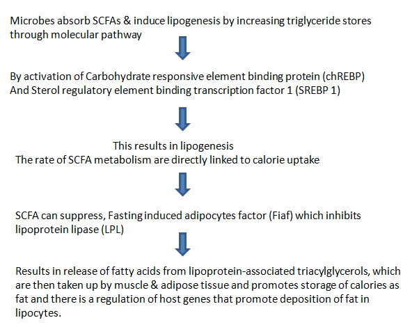
SHORT CHAIN FATTY ACIDS (SCFAs) AND OBESITY

Firmicutes breakdown carbohydrates in the gut, which can’t be digested by body’s enzymes such as dietary fibre & resistant starch, they ferment & produce metabolites, Short chain fatty acids(SCFAs),that are either absorbed by gut or excreted in feces.

CHRONIC SYSTEMIC INFLAMMATION AND OBESITY

On the basis of the recent demonstration that obesity and insulin resistance are associated with low-grade chronic
systemic inflammation, Cani et al postulated another mechanism linking the intestinal microbiota to the development of obesity. They hypothesized that bacterial lipopolysaccharide (LPS) derived from gram-negative bacteria residing in the gut microbiota acts as a triggering factor linking inflammation to high-fat diet–induced metabolic syndrome.

Gut microbiota also may contribute to metabolic disturbances observed in obese patients by triggering systemic inflammation. In a series of experiments mice fed with high-fat diet, they showed that (1) a high-fat diet increases endotoxemia and affects which bacterial populations are predominant in the intestinal microbiota, favour an increase in the gram-negative to gram-positive ratio), (2)chronic metabolic endotoxemia induces obesity, insulin resistance, and diabetes.

CONCLUSION

Certain metabolic products of gut microbiota, such as propionate & butyrate also play a vital role in maintaining proper body weight. Microbiome modulators (e.g. antimicrobials, diet, prebiotics or probiotics) mostly aimed to replace some of the defective microbes and specific commensal strains, probiotics, defined microbial communities, microbial-derived signalling molecules or metabolites helps to restore the healthy gut microbes by regulating normal metabolic process. Given the contribution of host genetics in many diseases associated with a dysbiotic microbiota, dual therapeutic strategies (e.g. combining immunotherapy and microbiota-targeted approaches) may also be required to restore the environment required to re-establish an effective communication between the host and the targeted microbiota. 

Reference

https://www.sciencedirect.com/science/article/abs/pii/S0025619611607027l

https://www.ncbi.nlm.nih.gov/pmc/articles/PMC2648620/

https://www.ncbi.nlm.nih.gov/pmc/articles/PMC5433529/

https://www.ncbi.nlm.nih.gov/pmc/articles/PMC5433529/

https://www.sciencedirect.com/science/article/pii/S1198743X14609769

ESTROGEN RECEPTOR SIGNALING IN BREAST CANCER

Our cells sends and receive millions of messages in the form of chemical signaling molecules. Cells communicate using these chemical signals. Regardless of nature of the signal, the target cell respond by the means of receptor, which binds the signal molecules(ligand) & initiates a response in target cell. Receptors are chemically protein or glycoprotein molecule which bind to signal molecules(ligand). Binding of ligand to its receptor causes conformational change in receptor that initiates a sequence of reaction leading to specific cellular responses. Based on location, Receptors are being classified as 1) Intracellular Receptors – Cytoplasmic Receptor & Nuclear Receptor 2) Cell Surface Receptors– G protein, Tyrosine Kinase Receptors & Janus Kinase Receptor

Estrogen and estrogen receptors (ERs) are critical regulators of breast epithelial cell proliferation, differentiation, and apoptosis. The Estrogen Receptors(ER) are believed to be a major contributing factor in the malignancy of breast cells. Targeting the ER signaling pathway has been a focal point in the development of breast cancer therapy. Approximately 70% of breast cancers are estrogen receptor-positive and this type of breast cancer is more common in postmenopausal women. Breast cancer, a genetically and clinically heterogeneous disease that originates from the mammary epithelial cells. It remains the leading cause of cancer deaths among females worldwide with about one in eight women (12 %) developing breast cancer in her lifetime. Woman’s risk for breast cancer is linked to her reproductive history and her lifetime hormonal exposure. The levels of estrogen in blood and tissue are associated with breast cancer carcinogenesis. Estrogen receptors belong to the Nuclear Receptor Superfamily. The two different forms of Estrogen Receptors(ER) – ERα  and  ERβ are coded by two distinct genes ESR1 and ESR2, located on chromosomes 6 and 14, respectively. ERα mediates unregulated cell proliferation in breast cells. However, ERβ opposes the actions of ERα by modulating the expression of ERα-regulated genes and reducing migration of cancer cells. Let’s know about Estrogen signalling pathway and what goes wrong in the pathway resulting in abnormal condition.

ESTROGEN SIGNALLING

Estrogen signaling includes two distinct pathways often referred to as genomic and non-genomic pathways. ERs are randomly distributed in the cell & are maintained in an inactive state by co-repressor proteins (NCORI, SMRT ). To activate ER- dependent transcription, ER-corepressor complexes need to be dissociated & replaced by co-activator complexes.

GENOMIC PATHWAY

From above figure- NR refers to Estrogen Receptor, Heat Stock protein refers Co-repressor

DYSREGULATION OF GENOMIC PATHWAY

Co- activators contain LXXLL motif (L,leucine, X, any A.A) that interacts with ligand binding domain of ERα. These co-regulators are associated with various enzymes such as deacetylase, acetyltransferase, methyltransferase, ATPase, etc which regulate chromatin remodeling & directly or indirectly regulate target gene expression. Deregulation of coregulator expression is associated with tumor progression, metastasis, cancer cell migration & drug resistance. Binding of AIB1 & BCAS3(Breast carcinoma amplified sequence 3)  both ERα co-activators are known to induce Breast carcinogenesis.

NON-GENOMIC PATHWAY

Estrogens regulate activity of ERs by serving as ligand, Estrone(E1), Estradiol(E2) &Estriol(E3)- 3 forms of estrogen. E2 is important ligand of ERs. Depending on a cell type, small fraction of ERα is involved in non-genomic action by anchoring to PM. Non genomic signaling of estrogen involves, mobilization of secondary messengers & interaction with membrane receptors such as, IGF-1R(Growth factor-1-receptor), EGFR(Epidermal growth factor receptor), Stimulation of effector molecules such as Src & PI3K, Serine/theronine protein kinase(AKT), MAPK(Mitogen activated protein kinase)

From above figure 1

B) Growth factors activate receptor tyrosine kinases, which activate MAPKs.  MAPKs can then phosphorylate and activate ERα either independent of E2 or synergize with E2 for optimal ERα activation.

C) Membrane associated ERα interacts and activates SRC kinase upon ligand binding. SRC kinase then phosphorylates and activates aromatase, which catalyzes conversion of androgens to estrogens within cells and amplify both genomic and nongenomic ERα-E2 signaling.

(D) Membrane anchored ERα can also activate various cytoplasmic kinases including PI3K-AKT-mTOR pathway through nongenomic actions, resulting in activation AKT pathway involved in cell proliferation and survival.

NORMAL REGULATED PATHWAY DEREGULATED PATHWAY
When E2 binds to activated ERα receptor , Depalmitoylation at cysteine477 (detachment of fatty acid molecule from the membrane)

The dissociated molecule then interacts with signaling molecule PI3K.


PI3K converts PIP2 into PIP3, which interacts with PH domain of AKT

Transfer AKT to cell membrane along with PDK1, resulting in conformational change in AKT & exposed T308 & S473 undergoes phosphorylation.

Further, PIP3-dependent phosphorylation by m-TOR, resulting in full activation of AKT/Protein Kinase B.

Promotes cell Proliferation, survival & apoptosis.
When E2 binds to activated membrane ERα, Palmitoylation at cysteine477 localize ERα to PM.

Responsible for ligand-induced activation of PI3K-AKT-mTOR pathway, resulting in Breast cancer cell.

ANTIBIOTIC RESISTANT BACTERIA

Bacteria are known to regulate their cooperative activities & physiological processes through a mechanism called “Quorum Sensing” (QS), in which bacteria communicate within themselves or within different species by releasing, sensing & responding to small diffusible signal molecules eg; Acylated homoserine lactones (AHLs) named as Autoinducer. QS allows microorganisms to control the behaviour of the entire bacterial population by secreting these signalling molecules. When the concentration of signal molecules reaches a certain threshold with bacterial population density, the expression of certain specific genes gets activated to regulate the bacteria population to undergo adaptation. These adaptations may activate a variety of cellular processes including virulence, drug resistance mechanism, tolerate antibiotics, biofilm formation in order to harm the host. QS exists in various bacteria, whether in Gram negative or Gram positive , but signal molecules they use to transmit information varies as different like human languages.

Antibiotics are used widely around the world besides antibiotic resistance is spreading faster than ever before. It takes 10-15 years to develop a new antibiotic but by this period bacteria become resistant to that drug, which is a serious problem to be considered. Most antibiotics currently used are designed to directly kill pathogenic bacteria. This “life or death” has caused them selection pressure promoting the evolution of microbial resistance and has become serious threats to human health. The large-scale use of antibiotics in the clinical treatment had led to multi-resistance mechanisms by microorganisms against the target of antibiotics.

Microbial Resistance mechanisms

1. Passivation of antibiotics through the chemical modification

Microorganism produces certain chemicals that inactivate the mechanism of the drug. By secretion of a modified enzyme changes the chemical structure of antibiotic drugs leading to inactivation or loss of activity of antibiotics. The enzymatic mechanism includes antibiotic degradation and derivatization of antibiotic chemical groups which leads to inactivating the drugs or destroying the molecules themselves so that no longer antibiotics can interact with microorganisms. eg; Bacterial enzyme, Beta-lactamase destroys the active component(the beta-lactam ring) of penicillins.

2. Efflux pump systemic

Here microorganisms use antibiotic pumps to expel antibiotics. Antibiotics usually enter the cell through the cell membrane in order to effectively attack specific targets. Microorganisms assemble efflux pump protein on their cell membrane to expel antibiotic drugs from cells. Bacterial efflux pumps are composed of the outer membrane channel protein, the fusion protein, and cytoplasmic efflux protein. Any foreign particle including protein, antibiotics is selectively or non-selectively eliminated from bacteria. Excretion rate is usually faster than drug penetration rate, thereby controlling drug level in the cell to non-sensitive level. They even have a variety of microbial efflux pump system such as lipophilic, hydrophilic efflux system that targets drugs of different chemical properties.

3. Modification of drug trageting genes

Microbes have evolved different strategies, which include modifying the target site, thereby reducing the affinity for antibiotic molecules to bind to the target site.

4. Biofilm Formation

Biofilm is an effective drug barrier, which significantly reduces antibiotic permeability. Bacteria are connected to each other through proteins & DNA, especially extracellular polysaccharides forming insurmountable barriers, which can reduce the permeability of antibiotic drugs & improve the survival rate of bacteria in the biofilm. Biofilms provide protection to microorganisms for altered PH, osmolarity, nutrients, mechanical or shear forces & also blocks access to antibiotics and the host’s immune cells.

This special environment in the biofilm makes bacteria in the membrane produce heterogeneity & produce antibiotics against other bacteria. Heterogeneity of bacteria has led to different levels of drug resistance against bacterial cells, where new research is emphasized more in this area.

It is essential for microbes to adapt & cope with these stressful situations. Thus a bacteria have devised complex mechanisms. Multiple drug resistance mechanisms expressed by these microorganisms had made it difficult to overcome and solve the problem of microbial resistance. In the struggle b/w microorganisms & antibiotics, more & more microorganisms have evolved multiple resistance mechanisms & have become “super bacteria”. Currently inhibiting bacterial QS has become a new promising antibacterial strategy.

Coumarin’s showed a Anti-Quorum Sensing Activity

Coumarins are a class of natural compounds based on benzopyrones. Coumarins are identified as secondary metabolites in about 150 different plant species. 7,8-dihydroxy-4-methylcoumarin was identified as the most effective anti-QS compound which had the ability to inhibit cell-to-cell communication in bacterial communities. At the same time when we look at its structural features, particularly the hydroxy group positions, well-responded to all anti-QS active coumarins. It was significantly noted as an increase in the antibacterial effect upon the hydroxylation of coumarins at positions 6,7, or 8. It showed that hydroxylation at position 7 increased anti-QS activity, while hydroxylation of coumarin at positions 6 & 7 decreased this activity in comparison to conventional coumarin. It also showed that Coumarin when combined with other Plant-Derived small molecules showed an effective response. 7.8-dihydroxy-4-methylcoumarin with 4-hexyl-1.3-benzendiol and gamma-octalactone demonstrated a synergetic anti-QS effect. Coumarins are characterized by a special mechanism through inhibition of the metabolism of cyclic 3,5-diguanylate (c-di-GMP), an intracellular intermediate involved in the regulation of bacterial exopolysaccharide synthesis, biofilm formation, adhesion, and virulence. Besides Coumarins are proposed for medical uses due to their proven biological activity such as Anti-inflammatory, Anti-ulcerogenic, Anticoagulant & are also Antioxidants.

Ref;https://www.ncbi.nlm.nih.gov/pmc/articles/PMC7143945/ ; https://www.mdpi.com/2076-2607/8/3/425/htm

YEAST PROBIOTICS IN THE TREATMENT OF IBD MOUSE MODEL

Inflammatory Bowel Disease (IBD), is a chronic and life-threatening inflammatory disorder of the gastrointestinal tract that arises due to host-microbial interactions in a genetically susceptible individual. It can affect people of all ages, including children & geriatric populations, and can affect all aspects of life. However, IBD is increasing in the emerging population, mainly in the industrialized countries of Asia, Africa, and South America. IBD is a group of autoimmune diseases that are characterized by inflammation of both the small & large intestine in which elements of the digestive system are attacked by the body’s own immune system. IBD includes Crohn’s disease & Ulcerative Colitis.

Crohn’s Disease; Is an IBD that causes inflammation anywhere along the lining of the digestive tract, from mouth to anus in a discontinuous pattern. Crohn’s exhibits histologically a thickened submucosa, transmural inflammation, fissuring ulceration & granulomas.

Ulcerative colitis; Involves the rectum & can affect part of the colon or the entire colon in a continuous pattern. Here the inflammation is limited to the mucosa & submucosa with cryptitis & crypt abscesses.

Causes; The exact cause of IBD remains unknown, one possible cause is an immune system malfunction, abnormal immune response causes the immune system to attack the cells in the digestive tract.

  • Nucleotide-binding oligomerization domain 2 (NOD2) is the first gene found to be associated with increase in the susceptibility to Crohn’s disease, which is frequently mutated in patients with Crohn’s disease, occuring in around one-third of the patients.
  • Environmental factors to plays a important role in pathogenesis of IBD. Studies have shown that increased consumption of milk protein, animal protein, medium chain fatty acids, fast foods containing many fat & sugar rich components exacerbate the development of Crohn’s disease.
  • Smoking is another example of disease- specific modifier that seems to worsen Crohn’s disease. Smoking has been shown to affect cellular & humoral immune response & to promote colonic mucus production.
  • Other factors that influence the development of IBD, includes not limited to psychological stress, appendectomy is another independent risk factor for developing Crohn’s disease.

IBD appears as a result of abnormal host immune responses to the intestinal microbiota. The gastrointestinal tract of the human body is colonized by a vast range of microorganisms that numerically exceed host cells by around 10 times. The gut contains 1000-5000 different species from Firmicutes, Bacteroidetes, Proteobacteria & Actinobacteria. These microorganisms contain around 100-fold as many genes present in the human genome.

The immunological dysregulation in IBD is characterized by epithelial damage (abnormal mucus production, defective repair), expansion of inflammation-driven by intestinal flora & a large number of cells infiltrating into the lamina propria includes T cells, B cells, macrophages, neutrophils results in failure of immune regulation. The intestinal immune system is divided into innate immunity & adaptive immunity. Innate immunity includes the barrier function of the intestinal mucosa, antibacterial proteins, the acid PH value of the stomach to limit microbial growth. Whereas Adaptive immunity is pathogen-specific & is usually initiated under the circumstances in which the innate immune responses cannot circumvent the stimulation of a pathogen. After exposure to a pathogen, it usually takes several days to finally activate adaptive immune responses, including T & B cells. The initiation of immune response to intestinal flora is tightly regulated & this regulation determines the occurrence of immune tolerance or a defensive inflammatory response. Disturbance of the balance of these responses can cause IBD.

What if we had a supplement that could sense the disturbance to the microbiome & restore balance to treat this chronic disease?

Francisco Quintana, an immunologist & neurologist at MIT’s Broad Institute & Harvard Medical school, and his colleagues have genetically engineered yeast that can respond to inflammation & successfully relieve IBD symptoms in mice. Previous studies have found that people with IBD have an unbalanced gut microbiome with lower overall diversity & more bacterial species that drive inflammation. Scientists believe that these yeast probiotics could help to restore this balance, neutralize & reduce the inflammatory molecule. Patients with IBD carry an abundance of microbial species that produces excessive ATP, which promotes inflammation. Meanwhile, host immune cells that drive inflammation also secrete ATP, further exacerbating the situation.

The researchers engineered Saccharomyces cerevisiae, the same yeast species that facilitate bread and beer-making, to detect ATP with the human receptors. They used CRISPR-Cas 9, a type of gene-editing technology, to introduce a gene that is activated by inflammation in the gut. They linked the activation of this receptor to secret the enzyme apyrase, which degrades ATP. The result is, probiotic that senses a pro-inflammatory molecule, extracellular ATP(eATP), generates a proportional self-regulated response to neutralize it. The engineered yeast produces different amounts of enzyme, depending on how many inflammatory signals are produced. In the mouse model of IBD, the probiotic performed better than standard therapies for IBD. This approach could work to mitigate inflammation not only in gut but also in other parts of the body. The therapy also helped to restore the balance of gut flora in the mice and this therapy can even help with other inflammatory conditions, including diseases like Parkinson’s disease that are anchored or initiated in the gut.

Saccharomyces cerevisiae

This approach is the next step of personalized medicine, going one step beyond that to adjust therapy only to those specific areas of the patient that are diseased. With this knowledge, we have the ability to develop novel personalized treatments for IBD patients.

Ref; https://www.the-scientist.com/sponsored-article/engineered-yeast-robots-destroy-inflammatory-molecules-in-an-ibd-mouse-model-69092

CORONAVIRUS : Inside the body

The Coronavirus disease 19 (COVID-19) is a highly transmittable & pathogenic viral infection caused by Severe acute respiratory syndrome coronavirus 2 (SARS-COV-2) which caused global pandemic that led to dramatic loss of human lives. Coronavirus can be classified into 4 genera namely; Alpha coronavirus, Beta coronavirus, Gamma coronavirus, and Delta coronavirus. When we look at its morphology it is large, enveloped, single stranded positive sense RNA virus with 5′ cap structure and 3′ Poly A tail structure & genome ranges from 27 to 32kb. It encodes 4 structural proteins on its surface, namely Nucleocapsid(N) protein, Membrane(M) protein, Spike(S) protein and Envelop(E) protein & several non-structural proteins. The genome is packed inside a helical capsid formed by nucleocapsid protein (N). The membrane protein(M) & envelope protein (E) are involved in virus assembly, whereas spike protein(S) mediates virus entry into host cells. The genetic material of the virus, RNA is made up of molecules, each part of this code contain instructions for how to make one specific protein that allows the virus to function. A virus has one goal, to make more of itself because it’s so simple it can’t do by its own, so it uses us. Every time virus infects a person it uses their cells to make copies of itself, while replicating this code again & again it eventually makes a mistake, sometimes it deletes or adds a letter, that mistake is called mutation and it slightly changes instructions for making the virus & that slightly altered virus is a variant.

ACCESS TO THE HUMAN CELLS

Virus has envelope containing spike protein on its surface. This spike protein on virus unlocks/ gains access to interior of human cell through ACE2( angiotensin converting enzyme2). Virus uses ACE2 as receptor to unlock the human cells. It has not been entry point for just coronavirus, but also for multiple virus. ACE2 receptor exists abundance on the surface of lung cells.

There is another second protein called TMPRSS2 which allows virus to reproduce & eventually leads it to transmission within the cell. It was found that cells from nasal cavity, eye, lung, gut, heart and kidney have these proteins. Now the virus overcomes the cell metabolism to its own advantage and uses cells to multiply & make its own replicas, so within 2 days you have millions of viruses in your body. Now your body starts recognizing this invasion, body then fights by producing antibodies which recognizes foreign virus, attacks them and kills them. This is the normal process that takes place in the body when any foreign bodies attacks.

Now within 4-5 days, when antibodies are sufficient in number, viruses are large in number there is fight between these two(antibodies & viruses). Our immune system now produces certain chemicals into bloodstream know as CYTOKINES these are chemicals that causes the body to have higher temperature, bodyache, like incase if you have throat infection, you experience sore throat this is because, cells in the throat have maximum of viruses & there is a fight going on inside and hence because of the irritation you get cough.

Incubation period for virus means, from the entry of the virus to its first symptom, on an average of 5 days. 1st symptom maybe fever, bodyache, headache, even diarrhea and has to be kept in the mind as first day of infection producing symptom. If fever goes higher or sore throat persist for 3 continuous days and on 4 -5 day if your fever comes down and you have no symptoms, check for another 5 days & if you have no symptoms, then you are a Corona Warrior. 80% of the people have only minor symptoms which settles in 3-4 days. Remaining 10-20% people takes dangerous turn. Fever which had settled within 4-5days, it comes back or if it was there now you have a higher degree of fever & if you had a cough, now you experience deep hacking cough. The 3 symptoms indicating that your disease is taking to a dangerous turn are;

  • Higher fever than before or if you have no fever, you will have new fever with higher temperature
  • Chest congestion or difficulty in inspiration & expiration process.
  • Deep hacking cough, which makes you difficult even when you are sleeping.

CYTOKINE STORM

When a foreign invaders enters our body, our immune system produces antibodies to destroy them. But sometimes our immune system gets hyperactivated & instead of killing foreign invaders, it gets excited itself & start killing its own cells, leading to cytokine storm.

Cytokines are the small proteins produced by the immune system, which act as signaling molecules that convey the message between different immune system cells. Your immune cells uses cytokines to communicate. Cytokine proteins are important in order to have unified immune response against foreign invaders. These signaling molecules are produced by many different immune cells, such as Mast cells, macrophages, B-cells, T- cells. There are few types of cytokines namely

Interferons – These protein inhibit virus from replicating. If a cell gets invaded by a virus, this signals other cells to put a shields so that virus doesn’t spread.

Interleukins- These protein regulate immune and inflammatory responses. They are mainly produced by white blood cells & their job is to send signals to WBCs to report for duty.

Chemokines- These call the cells to the site of infection. The ability to call in other cells using chemical message is a process referred to as chemotaxis.

Now if we get infected with virus or bacteria, the cytokine concentration increases and due to hyperactivated response of immune system, it releases more cytokines to signal neighboring immune cells & this leads to fail in identifying/differentiating between invader and our own cells.

CYTOKINE STORM EFFECTS

When virus attacks lung cell, they cause damage to cell. The damaged cells releases chemicals including histamine, prostaglandins etc .These chemicals sends signals to WBCs to recover from damage & to releases cytokine. When cytokines comes in the close proximity to the lung cells, there will be a huge rush of cytokines, signaling more & more WBCs to come in, now this area results in swelling. As more & more cytokines are moving in the close proximity to damaged lung cell, the wall of blood vessels becomes thinner and thinner, this causes leak of fluid from blood vessels into neighbouring lung tissue & as fluid is moving to lung tissue, now water content from blood vessels are moving out leaving with only a cellular components in blood vessel and due to this reason, blood cells in the blood vessels start sticking instead of free flow, leading to blood clot and when there is a clot formation, the required amount of blood, oxygen won’t reach to your different organs & hence Cytokine storm leads to Multiple Organ Dsyfunction. That’s the reason why most of the covid patient faced comorbidity death like liver failure, kidney failure, heart failure which is the part of cytokine storm.

DIAGNOSIS OF STORM

When our own cells have turned against us. We need to prevent release of cytokines in the body, we need to suppress our immune system. There are certain factors/ indications which has to be checked to prevent cytokine storm.

  • Ferritin protein; Increases high before the cytokine storm.
  • C- Reactive Protein(CRP) ; Increases high, not only here but in any sort of infection in the body.
  • Interleukin 6 ; Increases high, in the case of viral infection.

WHY FIRST LUNG CELLS ?

Virus is recognized by human body by our immune system, spike protein on the surface of virus is recognized by antibodies. Some cells in our body have similar appearance & structure to that of virus spike protein, these are specifically seen more in the blood vessels of lungs .

In the lungs, antibodies which are supposed to only attack foreign viruses, recognizes these similar cells & attacks them, causing normal cells to damage. when the cells get damaged, the normal smoothness of blood vessels interior is lost, cells lining the wall of blood vessels become rough hence blood cells start sticking there, resulting in formation of clot & blocks the passage of blood vessel and no more blood can pass. So if this happens in lungs, the normal exchange of oxygen that happens b/w air and your blood in the alveoli of your lung is not going to happen, no blood flow, no oxygenation and when no oxygenation your saturation of oxygen in your blood keeps going down, you start feeling breathlessness & have a problem in breathing depending upon the severity of damage to the lungs. This is all not because of virus persisting in your lung but because body’s reaction to the viruses, autoantibodies attacking our own cells.

ROLE OF STEROIDS

On 5th or 6th day if patient has come up with high fever, bad cough & breathlessness, saturation below 94% this is where steroids has to be given to prevent cytokine storm and another important thing is, steroid alone doesn’t stop the clotting of blood within the lung blood vessel, so we have to combine it with anticoagulant. The 2 medication which has to the taken at this stage, steroids with anticoagulant. In the case of steroids, timing is very important & taking steroids all alone without the consultation is again a dangerous thing. Taking steroids at the early stage of disease ( first 5 days of the disease), results in multiplication phase of the virus to be prolonged, leading your disease to be prolonged.

Steroids are used to treat inflammatory conditions which may be seen in serious COVID-19 cases. WHO recommends corticosteroids (i.e dexamethasone, hydrocortisone or prednisone) to give orally or intravenously for the treatment of patients with severe COVID-19. Treatment showed to reduce the length of time on ventilation for the patients with critically ill. Steroids has to be taken once a day for atleast 7-10 days & anticoagulants has to be continued for 3 weeks, not 10 days like steroid because tendency of blood clotting has 3 weeks, so that it can cover. Dosage of the steroid shouldn’t be more than 2mg/kg. Though steroids are effective to reduce infection associated with inflammation but they suppress our immunity. When you are on the course of steroids & anticoagulants dramatically you become better, fever is gone, cough is better, but again on 5th day on the course of steroids, you start having a fever again and this time it is not virus, but as normal immunity is being suppressed, a normal bacteria in your lungs attacks your body and causes Pneumonia which should be covered with antibiotics. Steroids do benefit in huge way in life saving way, but it encourage infection by bacteria within our lungs and this is the only stage, when antibiotics has to be taken. Taking antibiotics at the 1st symptom is actually encouraging bacterial resistance, resulting in bacteria becoming resistant against drug, because no antibiotics ( like Azithromycin or Doxycycline) do anything in eliminating the virus, they doesn’t work against virus But are useful in protecting patient from bacterial pneumonia which happens at the 5 day after starting steroids. Steroids are diabetogenic- those who are pre-diabetic they become frank diabetic, when they are on steroids which is the another drawback of steroids.

Than taking our disease to more complications and to the dangerous turn, let’s prevent it and it is important to take right medication at the right time and to follow the simple protocols, such as wearing a mask, social distancing, washing hands. The novel coronavirus spreads so rapidly that it has changed rhythm of the globe. SARS-Cov-2 virus entered our country in March 2020 and somehow we managed to come out of first wave. It again strike our nation in its mutant form & thus Covid 19 second wave emerged. Now again epidemiologists and various scientists have predicted 3rd wave. So let us together prevent it.

Ref;https://youtu.be/CJzo6JIqhCw

MICROBES IN PLASTIC WASTE TREATMENT

Humans have always created waste over a years. Solid waste, garbage, trash has been dropped into streets, sewage, landfills, waterways all over. Advances have been made globally to confront complex problems of waste generation, collection, and disposal. Currently, about 2.01 billion metric tons of municipal solid wastes are produced annually worldwide. The world bank estimates, overall waste generation will increase to around 3.40 billion metric tons by 2025. Out of total municipal waste collected on average, 94% is dumped on land and 5% is composted.

Nowadays, Plastics have become a worldwide recognized problem. These synthetic durable, non-reactive, cheap, conventional plastics play a vital role in our day-to-day life. They have taken constant place in our lives, by providing series of incredible benefits, besides they bring huge remarkable impact on soil, on our environment and health, Humans have generated around 380 million tons plastics every year, from which only 9% has been recycled and remaining ends up in landfill sites or oceans and recycled plastics causes more problems to the environment than original plastics due to the mixing of additives, colors, stabilizers, etc. Hence, there is an urgent need of reducing mankind’s dependencies on these nonrenewable resources. To overcome these crises, Biodegradable plastics take the best option to protect the environment against petroleum-based plastics.

There are many biodegradable plastics with different degrees of biodegradability such as starch-based, soy-based, cellulose-based, lignin-based, but among them, Polyhydroxybutyrate( PHBs), bacterial based bioplastics are the best and 100% biodegradable ones. PHB is a biologically produced biodegradable substance synthesized as inclusion bodies in numerous bacteria like Bacillus, Azotobacter chroococcum, Ralstonia eutrophus, Bacillus megaterium etc. PHBs are the energy reserve in the form of water-insoluble dense granules present in the cytoplasm of bacteria, they use them to store energy when they lack enough nutrients to reproduce. When microbes are under stress conditions or imbalanced condition of nutrients or in presence of excess carbon sources, they result in the production of granules inside their cells, resembling a very good property to those of synthetic plastics besides fully biodegradable. On degradation, they produce carbon dioxide, methane, mineral salts, water, and biomass to the environment and this PHB is well-posed for commercial adoption use due to better biodegradable properties in many types of environment.

Through these kinds of an alternative approach for the production of renewable, biodegradable plastics, could make a huge difference in the treatment of waste management, PHB is widely used for various biomedical applications, food packaging, plant pots, packing of beauty products, pharmaceutical industries, etc. Further chemical modifications with PHB could result in properties of the polymer to great extent. Thus by increasing the production of PHB, could bring a positive environmental effect. In next coming years, statistics of plastic waste usage could drastically reduce to make a better, toxic-free, green environment.

HONEYBEE’S VENOM AS A LIFE SAVING DRUG

Honeybee venom, a weapon against the Breast cancer.

As we are already aware that Cancer is one of the most adaptive and life-threatening enemies. If we look at 2020 statistics, breast cancer patients have greatly increased in recent years about 2.26 million cases worldwide. The 5-year survival rate for breast cancer patients has greatly increased in recent years and is currently about 90%. About 1 in 28 women are likely to develop breast cancer during their lifetime. and about only 1 in 8 women will be diagnosed with breast cancer and about 1 in 39 women die from this disease. Breast cancer is one of the most common forms of cancer affecting women around the world. Breast cancer is a disease in which cells in the breast grow continuously out of control. Breast cancer has become the second most common cause of cancer-related death in countries. The exact cause of this breast cancer is unknown. However several factors affect the risk Of developing breast cancer. The chances of developing breast cancer may depend on the combination of genes, our lifestyle, habit, our choice, and even the environment in which we dwell.

Some of Risk factors affecting the Breast Cancer.

  1. AGE

The risk of developing breast cancer increases with age. About 8 out of 10 cases of breast cancer happen with women over around age of 50 who have been through menopause. All women around 50 to 70 years of age should be screened for breast cancer every 3 years as part of the NHS breast screening Programme.

2. HORMONES

The female hormone estrogen can sometimes stimulate breast cancer cells and can cause them to grow. The risk of developing breast cancer may rise slightly with the amount of estrogen your body is exposed to. For example, if you start menarche at a young age of 12 and experience menopause later than average of around 55, therefore you will have been exposed to estrogen over a longer period of time and in the same way, not having children or having children at later in life might slightly develop the risk of breast cancer.

3 . GENETIC MUTATIONS

Inherited changes, such as mutations to certain genes, such as BRCA1 and BRCA2.Women who have inherited these genetic changes are at higher risk of breast cancer.

LIFESTYLE FACTORS.

  1. BEING OBESE OR OVERWEIGHT

If you have experienced the menopause and are overweight, you may have slightly the risk of developing the breast cancer.

2. ALCOHOL

Drinking alcohol increases the risk of getting breast cancer.

3. RADIATION

Women who had experienced radiation therapy like treatment for Hodgkin’s lymphoma, before the age of 30 have a risk of getting breast cancer in their later stages of life.

These are some of the risk factors known to affect your likelihood of developing breast cancer. Some of these you can’t do anything about, but you can help lower your risk of breast cancer by taking care of your health by keeping a healthy weight, regular exercise changing your life habits etc.

Some of the Cancer subtypes such as Triple negative and HER2-enriched breast cancer accounts for disproportionate for number of deaths in women with this disease. This triple negative breast cancer is one of the most aggressive form and hard to treat type of forms . It accounts for 10-15% of all the breast cancer. Treating this Triple negative and HER2 involves chemotherapy, radiation and surgery. As we are already aware that these medication could kill most of normal cells and have less results. Hence currently there are no clinically effective targeted therapies for this. Thus the survival rates are less with Triple negative breast cancer.

But, a new outbreaking study found that venom from honeybee could destroy these aggressive and resistant breast cancer cells. Harry Perkins Institute of Medical Research in Western Australia found that honeybee venom and its active component is known as melittin could kill these aggressive and hard to treat a type of cancer, with negligible effects on normal cells. Bee venom has the extraordinary property of anticancer. Researchers said honeybee venom has proven to be extremely potential at concentrations that are not as damaging to normal cells. Melittin is a naturally occurring substance in honeybee venom. It is the molecule that creates the painful sensation of a bee’s sting. This compound on its own was found to be effective in disrupting the cancer cells’ growth. This Melittin compound even acts on a wide range of tumors including lung, ovarian and pancreatic cancers.

How it actually worked ?

The study showed a specific concentration of venom killed 100% of triple-negative breast cancer and HER2 -enriched breast cancer cells. For the study, bees were put to sleep with Carbon dioxide. Later they were kept on ice before the venom was extracted and injected into tumors. When they injected, they observed Melittin impedes the chemical messages that trigger signaling pathways within breast cancer cells to reduce cell growth and replication all within 20 minutes. Melittin modulated the signaling in breast cancer cells by suppressing the activation of receptor that is commonly overexpressed in triple-negative breast cancer. Scientists produced Melittin synthetically in labs. They combined it with existing chemotherapy drugs such as Docetaxel. This Melittin compound enters the surface of the cell or plasma membrane and forms pores causing cells to die. This result was achieved within 60 minutes. The venom had very minimal effect on normal healthy cells. As result, it worked effectively in reducing tumor growth cells in mice and they hope the discovery could lead to the development of treatment for triple-negative breast cancer. So that, humans can live forever and healthier life.

THUS, this provides a wonderful example of where compounds in nature can be used to treat human diseases to solve real-world problems. The natural product could be efficacious sources for the prevention and treatment of many diseases. These natural products from herbal remedies, medicinal plants, and their constituents have been used to treat various diseases including cancer from ancient times. Our Nature has the art of healing and it is an essential part of our health and survival. So let us adopt the pace of nature without harming her.

Ref ; http://www.nhs.uk ,http://www.cdc.gov , http://www.healthline.com ,http://www.breastcancer.org

IMMORTAL JELLY FISH

Turritopsis nutricula (The immortal jelly fish)

There is organism in great wide world of ours ,Turritopsis nutricula defining itself as an immortal organism in animal kingdom.

Distribution : Originating from the Caribbean sea ,this unique creature has spread all over the world and now it has been found in both temperate and tropical regions,though it prefers warmer water.

SIZE : It is around 4-5 millimeter tall and wide.

This Immortal jelly fish has ability to reactivate its genetic instructions from earlier in its life cycle.Thus by escaping death it achieves potential immortality. In case of sure death,it revert back to young version of itself.

Now, let us try to understand its life cycle

Usually corals, sea anemones & jelly fish belongs to group of animals called cnidarians. Turritopsis nutricula belongs to hydrozoans. Some jelly fish change forms at different phases of their life cycle,while others remain in one form for their entire life.There are two basic body shapes which exit in the life cycle of jelly fish.

  1. Polyp form ( which remains attached to surface)
  2. Medusa form (which is a free floating,adult form)

Now let us the discuss the way that they are biologically immortal. Normally ,in hydrozoans life cycle ,Medusae ( matured jellyfish) spawn( fertilize) sperm and eggs. Any fertilized eggs becomes small larvae called PLANULA. when these planula settles somewhere below the sea,a new hydroid colony grows once again which are called as POLYPS ,these eventually grow in colonies and releases more medusae and these medusae develop into matured jellyfish and this is how the cycle is with both sexually and asexually reproduction.

However, Turritopsis are different.when they become threatened by physical activities or starvation , in such conditions they transform back to juvenile polyp stage by shinking their tentacles and body they sink to ocean floor to start a new cycle . over a time when a condition is favorable, new genetically identical clones of original jellyfish are released from polyp into sea and later polyp gets transformed into medusae and later medusae gets mature and develop into mature jellyfish and again if they are threatened ,the process can be repeated over and over. The way T.nutricula is able to live forever is through the process called as TRANSDIFFERENTIATION,it is process in which change of cell or tissue from one differentiated state to another OR it is the process of differentiation of a tissue-specific stem cell into another type of cell,for example,a bone marrow stem cell differentiating into a neuron.During this process it help them to survive by reducing metabolic requirements which help them to turn to polyp besides it can encyst. As a cyst T.nutricula can stay dormant for extended periods of time until environmental conditions are favorable to restart its life cycle. In most of jellyfish,once their adulthood is reached, asexual reproduction is no longer possible. But T.nutricula has two forms. Sexual reproduction and asexual reproduction, if there are enough sexually mature jellyfish, will be able to reproduce sexually and if, population density is low it can just revert back and can produce asexually,which is known as biologically immortal.

BENIFITS OF T.nutricula

THIS natural process( Transdifferentiation)has inspired many researchers to find stem cells to renew damaged or dead tissues in humans,so that many diseases like cancer and many more can be treated. This organism has found itself as great important. If a cell or organism undergoes aging , there are two biological processes. 1. Negative regulation of cell division. 2.Positive regulation of apoptosis may occur. Furthermore aging and neurodegenerative diseases are directly associated with these two biological processes while cancer ,aging does not happen hence these two processes are inversely associated. Thus T. nutricula has been a great tool in cancer ,aging and neurodegenerative related disorder.

Another interesting thing is these soft bodies rely on thousands of venom containing stinging cells on their tentacles called Cnidocytes, for their protection and prey capture. this venom ejected via a nematocyst , is a whip like a hollow tube which lies coiled under high osmotic pressure. when mechanical or chemical stimuli activate ( physical touch) ,an external trigger happens ,the lid of the cell breaks and opens,due to this sea water rushes in due to change in osmotic pressure ,this forces a microscopic barbed harpoon (nematocysts) to shoot out ,to penerate and inject into victim.

Humans might benefit from these stings of jellyfish, scientists are working on manipulating Cnidocytes to deliver medicine with nematocytes rarely 3% of size of typical syringe needle so it would help in the medical field. Besides it has harmful effects too ,the venom released by nematocysts causes contraction in heart muscles and even rapid death in case of rapid dose. Thus in such cases we should remove nematocyts by applying salt water followed by hot water and in these cases avoid applying cold water or fresh water.

One day in future humans may live for hundreds or even thousands of years and this may well be achieved because of research on immortal jellyfish . You could have ability to transform back into younger version of yourself enabling you to live forever.

REf:http://ed.ted.comhttps://youtu.be/yQduHyiWe9o ref:https://www.google.com/url?sa=t&source=web&rct=j&url=http://tnutricula-bethelu-corylundeen.weebly.com/unique-life-cycle.html&ved=2ahUKEwijt5-zoe_qAhWf4HMBHSrYCVYQFjAVegQIBRAB&usg=AOvVaw1m9nsa3uJn8oMawITwd_8h&cshid=1596183811583

BATS,THE FLYING MAMMALS

BATS

Bats are absolutely incredible animals.They are creatures of mammals that possess the extraordinary ability of powered flight.Bats are the smallest & fastest little creatures,reaching the speed of over 100 miles per hour.Despite their resemblance to rodents,bats are more closely related to humans than they are to mice.

Types of bats

There are 2 main types of bats namely,Microbats & Megabats . Most bats are Microbats which eat insects like moths,that come out at night.Vampire bats are only species of microbats that feed on blood rather than insects. But not to fear,they prefer blood of horses & cattles ,not humans.These bats don’t actually suck blood. Instead, they use their sharp teeth to small incision in skin of animals and then they consume blood as it runs from the wound.

Interesting fact is, Microbats rely on echolocate,also know as biosonar(biological sonars) by making a high pitched sound that travels until it hits an object & bounces back to them,which helps them to know about the object ‘s size & how far away is it. This ability is so sensitive that it can detect objects as thin as single strand of human hair.

In contrast ,these Megabats have larger eyes & stronger sense of smell than microbats but have smaller ears as they don’t echolocate.

MEGABATS

MICROBATS

DO YOU KNOW THESE FACTS ??…. Ever thought about these🤔

1.Why do bats hang upside down?

Hanging upside down for even few seconds will leave anyone feel dizzy and little light headed,then how do these bats do it??

Humans and bats circulatory system are fundamentally different,because our blood pumps in the direction of our brain,the force of gravity transfers even more blood to head when we hang upside down. Bats circulatory system pumps in opposite way,away from its head.They have a little pathways in their body through which blood passes,called arteries.These arteries have special valves in them that only let blood through one way.All mammals have valves in their veins that prevent blood from flowing backward, bats possess these valves in their arteries as well. All these adaptation ensures blood to distribute evenly throughout the body. Bats on other hand are tiny small compared to humans&they don’t weigh enough for gravity to affect their blood flow.

Bats have a good reason to hang upside down. As their wings aren’t as strong as birds wings, so they aren’t able to build up enough speed while taking off from ground or sitting position, Instead they can enter into fly with no effort if they start from hanging upside down. They take less effort to hang upside down, as they possess a specialized tendons in feet which enable them to hang in perfectly relaxed posture. But if they are sitting right side up they’d to contract muscle&thus expend energy to let go & begin flying

2.Bats are inspiring medical marvels

About 80 medicines come from plants that rely on bats for their survival.while bats are not blind ,studying how bats use echolocation has helped scientists develop navigational aids for blind and the peptides from bats are unusually selective in their mode of action,making them more therapeutically useful&this could potentially help doctor in the treatment of range of disorders featuring heightened pressure in small blood vessels or may be able to improve blood flow to damaged or transplanted tissue such as skin grafts.

3.Bats and the pollination&pest control

Over 300 species of fruit depend on bats for pollination.They help in spreading seeds for nuts,figs&cacao,without bats we could not have been imagined plants like agave or suguaro cactus.

Another benefit thing is there’s no need to use harmful pesticides when you have colony of bats nearby.Some individuals of bats can eat more than 600 flying insects per hour.Yes,we can perfectly choose bats for organic pest control, without them we humans might have faced huge problems towards our insect overlords.

4.Bats and their immune response

Bats,the only mammals which has ability to fly ,which take enormous output of energy as a byproduct of all metabiotic activity.This high production of energy,helps bats to tolerate against various kinds of viruses&other deadly diseases and this ability of production of high energy also can lead to accumulation of free radicals,a reactive molecule that can damage tissues.To prevent this reactive waste from damaging bats have sophisticated defence mechanisms,which even helps them to defend themselves from deadly viruses&diseases.

Do you know why bats carry so many diseases??

To have a clear idea about this,let’s have a view on this below video

https://youtu.be/Ao0dqJvH4a0

https://ed.ted.com/best_of_web/CnfJm2rX#review

Gene Editing

What is Gene Editing?

Genome editing, or genome engineering, or gene editing, is a type of genetic engineering in which DNA is inserted, deleted, modified or replaced in the genome of a living organism.

Hand of scientist replacing DNA – genetic engineering and gene manipulation concept

Rather than directions going one way from DNA to RNA to proteins,latest study shows that RNA itself modulates how DNA is transcribed using Gene Editing

Genome editing is technology that give the ability to change organisms DNA or can be altered at particular locations in genome.CRISPR-CAS9 is a recent genome editing which has been developed.It is short clustered regularly interspaced short palindromic repeats & is associated with protein 9.

CRISPR -CAS9 System consist of two components

  1. DNA cutting protein called CAS9.
  2. RNA molecule known as GUIDE RNA (sg RNA) bound together,they form a complex,that can identify & cut specific sections of DNA

IDEA BEHIND THE CRISPR-CAS9

CRISPR-CAS9 was adapted from naturally genome editing system in bacteria.The bacteria capture snippets of DNA from invading viruses & use them to create DNA segments known as CRISPR arrays.The CRISPR arrays allow the bacteria to ”remember” the viruses.If viruses attack again,bacteria produces RNA segments from CRISPR arrays to traget the viruses DNA.The bacteria then uses CAS9 enzyme to cut particular part of DNA.

How Does CRISPR -CAS9 Works?

CRISPR -CAS9 work similarly,A small piece of RNA with short ‘Guide’sequence that attaches to target sequence of DNA in genome.RNA also binds to CAS9 enzyme .As in bacteria,the modified RNA is used to recognize the DNA sequence,CAS9 enzyme cuts the DNA at targeted location .Instead CAS9 other enzymes like(cpf 1) can also be used.Once DNA is cut,uses the cells own DNA repair machinery to add or delete pieces of genetic material or to alter DNA by replacing an existing segment with customized DNA sequence.

Now let us have a clear idea by having a view on this below video.

Recently,CRISPR gene editing in US for the first trial in cancer patient suggets that technique is Safe.